 SingaporeSG
 ChinaCN
 MalaysiaMY
 IndonesiaID
 MyanmarMM
Life Science
Quantitative Phase Imaging Holographic Monitor

HoloMonitor ® M4FL
When Holography meets Fluorescence
- Combining holography and fluorescence data in one experiment
- Minimal light exposure to reduce photocytotoxicity
- Small footprint so it fits inside your incubator
- Long-term, single-cell and cell population data
- Intuitive and guided workflow from setup to analysis
Unlock the Power of Fluorescence
HoloMonitor M4 + Fluorescence = HoloMonitor M4FL
The fluorescence add-on is compatible with the HoloMonitor M4 system. This way, you can tailor the HoloMonitor Live Cell Assays to your needs and add fluorescence to your HoloMonitor system to best fit your research application.
- Single Cell Tracking Assay
The HoloMonitor® Single Cell Tracking Assay enables automatic adherent cell tracking for detailed label-free studies of single-cell behavior with optional fluorescence.
- Kinetic Dose Response Assay
The label-free HoloMonitor® Kinetic Dose-Response Assay is designed for automated and detailed analysis of drug dose responses in adherent cells
- Kinetic Cell Motility Assay
The HoloMonitor® Kinetic Cell Motility Assay is ideal for studies investigating how different drug treatments influence cell population movement over time.
- Cell Morphology Assay
The HoloMonitor® Cell Morphology Assay enables high-content, non-invasive and kinetic analysis of a wide range of morphological properties with optional fluorescence.
- Wound Healing Assay
The HoloMonitor® Wound Healing Assay enables automatic, non-invasive cell migration analysis giving you accurate, kinetic gap closure data in real-time.
- Cell Quality Control Assay
The HoloMonitor® cell culture quality control (Cell QC) assay ensures your cells’ quality, avoids contaminated cells, and detects undesired cell changes.
- Cell Counter
The HoloMonitor® Cell Counter is designed for automated cell counting of suspension cells with high-precision results within one minute.
- Kinetic Cell Proliferation Assay
HoloMonitor® Kinetic Cell Proliferation Assay is designed for automated and detailed proliferation analysis of adherent cells.
App Suite 4.0 software for fluorescence
The HoloMonitor® App Suite cell imaging software facilitates kinetic live-cell tracking and analysis of your cells right from your incubator. In addition to detailed single-cell and cell population data, App Suite provides you with visual data in the form of high-quality images and time-lapse videos of your in vitro cell cultures. Moreover, you can easily re-analyze the imaging results of previous experiments. With the HoloMonitor live cell assays, you can extract multiple results anytime from your samples. And, of course, this cell imaging software allows for convenient data analysis from outside the lab.
Featured Applications
Illuminate, Analyze and Discover
- Cell Death
- Reporter Gene Expression
- Co-Culture
- Transfection Efficiency
- Uptake Assay
- Live Cell Staining
Assay Output
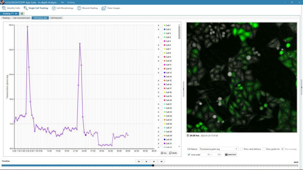
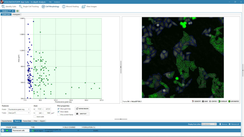
In addition to all the label-free holography cell features to study your cells’ behavior, you can analyze your cells’ fluorescence signal in HoloMonitor’s Single Cell Tracking and Cell Morphology Assay.
Single Cell Tracking Assay
The HoloMonitor® Single Cell Tracking assay offers user-friendly software for continuous, label-free imaging and tracking of adherent cells, enabling in-depth characterization of heterogeneous cell behavior at the single-cell level. With the add-on fluorescence unit, you can add a green fluorescence dimension to the holography data, providing enhanced insights into your cell cultures.
Cell Morphology Assay
The label-free HoloMonitor® Cell Morphology Assay enables non-invasive live cell imaging and kinetic analysis of a wide range of morphological properties, including individual cell volume, area, and thickness. With the add-on fluorescence unit, you can add a green fluorescence dimension to the holography data, providing enhanced insights into your cell cultures.
HoloMonitor M4FL User Reviews
HoloMonitor M4FL is on the trusted Select Science review website. You can explore experiences from other scientists about our live cell analysis tools, their various applications in cell research, and our services.
Read more reviews HERE.
| 1-channel fluorescence | Optimized for EGFP (FITC-Cy2) |
| Excitation | 470 nm filtered LED, with bandwidth (FWHM): 40 nm |
| Emission | 525 nm filtered, with bandwidth (FWHM): 50 nm |
| Exposure time | 1 – 1000 ms |
| Gain | 1 – 4 |
| Magnification | 10x, matched with mounted HoloMonitor M4 |
| Sensor | 1280 x 1024 px, 1.31 MPix |
| Image Resolution | 0.5 µm |
| Field of view | 0.25 mm² |
| Image size | 1024 × 1024 pixels |
| Dimensions | 310 x 180 x 85 mm (W × D × H) |
| Weight | 4.0 kg |
| Power | < 5 W |
| Interface | USB connector |
| Software requirement | HoloMonitor App Suite software 4.0 |
| Operating environment | 10 – 50 °C, < 95 % relative humidity |

Authors: Frida Berlin
Journal: Doctoral thesis (2023)
Research Areas: Cell biology
Keywords: HoloMonitor M4, Mast cells, proteases, tryptase, chymase, chronic respiratory diseases, asthma
Chronic respiratory diseases, such as asthma, are an increaseing health issue worldwide and cause about 3.9 million deaths annually. Despite this, little is know about the molecular mechanisms underpinning disease pathogenesis. Bronchial and alveolar remodeling and impaired epithelial function are typical characteristics of chronic respiratory diseases. In these patients, an increased number of mast cells, positive for the serine proteases; tryptase and chymase, infiltrate the epithelium and the alveolar parenchyma. While it is likely that the epithelial cells are exposed to various amounts of released tryptase and chymase, the interaction between mast cells and epithelial cells remains unknown. This thesis aimed to investigate the impact of mast cell proteases on bronchial and alveolar remodelling. Human bronchial and alveolar epithelial cells were treated with tryptase and chymase. Holographic live cell imaging, fluorescent microscopy, and gene and protein assays were used to analyze various parameters such as proliferation patterns, protein expressions and distributions. The results showed that both tryptase and chymase promoted epithelial remodelling in several ways. Tryptase induced cell growth, cell survival, and wound healing, whereas chymase reduced cell growth, altered cell morphology and impaired epithelial barrier proprties. In conclusion, our results suggest that intraepithelial and alveolar mast cell release of proteases plays a crucial role in epithelial homeostasis, and that an inbalance of the protease release may be involved in respiratory disease progression and in disruption of critical epithelial functions.
Authors: Li et al.
Journal: Experimental Neurology (2023)
Research Areas: Cancer research
Cell Lines: U251, U373, HEK293T, HA
Keywords: HoloMonitor M4, cell motility, Glioblastoma STK24P1, P1-121aa, ELF2 Phosphorylation
Glioblastoma (GBM) is the most common malignant tumor of the central nervous system. Vasculogenic mimicry (VM) is a hematological system composed of tumor cells that exert blood perfusion without relying on vascular endothelial cells. The current poor results of anti-vascular therapy for clinical GBM are associated with the presence of VM; therefore, it is important to investigate VM formation in GBM. The results demonstrate thatSTK24P1 encodes P1-121aa with a kinase structural domain, and in vitro kinase assays demonstrated that P1-121aa mediates modification of ELF2 phosphorylation. ChIP and dual luciferase reporter gene assays demonstrated that the transcription factor ELF2 binds to VE-cadherin and the VEGFR2 promoter region, thereby promoting VM formation in glioma cells. P1-121aa, encoded by the pseudogene STK24P1, phosphorylates ELF2 at S107, increasing the stability of the ELF2 protein. ELF2 promotes VEGFR2 and VE-cadherin expression at the transcriptional level, which in turn promotes VM in GBM. This study demonstrates the important roles of STK24P1, P1-121aa, and ELF2 in regulating VM in GBM, which could provide potential targets for GBM treatment.HoloMonitor M4 is used to study the effect of different mutations on the migration ability of human glioma cell.
Authors: Wasilewska et al.
Journal: International Journal of Biological Macromolecules (2023)
Research Areas: Materials Science
Cell Lines: MC3T3-E1
Keywords: HoloMonitor M4, Cell proliferation, Cell morphology, Macroion multilayers, Polysaccharide layers, Label-free biosensors, Streaming potential, OWLS, Resonant waveguide grating, Cell adhesion, Antimicrobial coatings, QCM
The regulation of cellular adhesion is a crucial aspect in the development of biomaterials and cell-based biosensing assays. In this study, synthetic poly(diallyldimethylammonium chloride) (PDADMAC), natural chitosan, and heparin were utilized to assemble PDADMAC/heparin and chitosan/heparin films. The physicochemical properties of these macroion multilayers were characterized using streaming potential measurements (SPM), quartz crystal microbalance (QCM-D), and optical waveguide lightmode spectroscopy (OWLS), while their topography was imaged using atomic force microscopy (AFM). The adhesion of the preosteoblastic cell line MC3T3-E1 on these well-characterized polysaccharide-based multilayers was evaluated using a resonant waveguide grating (RWG) based optical biosensor and digital holographic microscopy HoloMonitor M4. Results showed that PDADMAC/heparin films were the most effective in inducing cellular adhesion, while chitosan/heparin-based multilayers exhibited negligible cell attachment. These findings suggest that polysaccharide-based multilayers have potential for use in medical applications.HoloMonitor M4 is used to study the cell proliferation and morphological parameters of preosteoblastic cells on different coatings.
Authors: Frida Berlin et al.
Journal: Cells (2023)
Research Areas: Cell Biology
Cell Lines: BECs, BEAS-2B, AECs, A549
Keywords: HoloMonitor M4, cell proliferation, mast cell, proteases, tryptase, bronchial epithelium, alveolar epithelium, growth factors, anti-apoptosis, airway remodeling, cell growth, par-2
The article investigates the role of mast cell tryptase in bronchial and alveolar remodeling and the mechanisms of regulation during inflammation. The study found that mast cell tryptase enhanced human bronchial and alveolar epithelial cell growth and shortened the cell division intervals. The elevated cell growth induced by tryptase remained in a pro-inflammatory state. Tryptase also increased the expression of the anti-apoptotic protein BIRC3, as well as growth factor release in epithelial cells1. HoloMonitor is used to investigate the cell growth and cell division effects of mast cell tryptase on bronchial and alveolar epithelial cells.