Magnetofection Transfection reagent - for a superior nucleic acid delivery in primary and hard-to-transfect cells
PolyMag NeoMagnetofection™ Reagent is specifically designed to achieve high transfection efficiency combined with superior transgene expression level. It is composed of magnetic nanoparticles coated with cationic molecules. PolyMag Neo is the ideal transfection reagent for a wide variety of cells.
High transgene expression
High transfection efficiency
Multipurpose (various types of nucleic acids)
Universal (primary cells and cell lines)
Simple, ready-to-use and rapid
Non toxic
Compatible with and without serum-containing culture media
Sizes:
100 µL: 100 transfections with 1µg of DNA
200 µL: 200 transfections with 1µg of DNA
1000 µL: 1000 transfections with 1µg of DNA
#KC30200 : Starting Kit with Super Magnetic plate + 100 µL of PolyMag Neo, PolyMag + CombiMag
#KC30296 : Starting Kit with plate 96 magnets + 100 µL of PolyMag Neo, PolyMag + CombiMag
#KC30400: Super Starting Kit with Super Magnetic plate + 100 µL of PolyMag, CombiMag, PolyMag Neo & 200 µL of SilenceMag
#KC30496 : Super Starting Kit with plate 96 magnets + 100 µL of PolyMag, CombiMag, PolyMag Neo & 200 µL of SilenceMag
Storage: +4°C
Shipping conditions: Room temperature
This reagent needs to be used with a magnetic plate
CATALOG NUMBER
UNIT SIZE
PG60100
100 µL
PG60200
200 µL
PG61000
1 mL
KC30200
Starting Kit
KC30296
Starting kit plate 96 magnets
KC30496
Super Starting Kit plate 96 magnets
KC30400
Super starting kit with Super Magnetic Plate
Applications
Suitable for all nucleic acids delivery: DNA, oligonucleotides, mRNA, siRNA, shRNA...
Improve your transgene expression experiments
Perfect for all transfections: Transient, stable, gene silencing, with or without serum
RECOMMENDED FOR: Transfection of primary and hard-to-transfect adherent cells
Results
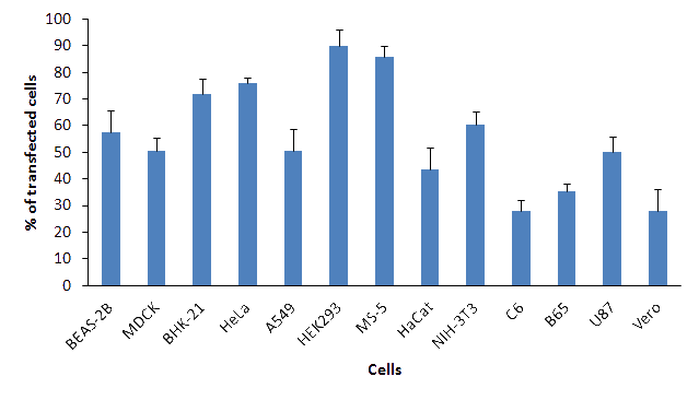
Figure 1: PolyMag Neo transfection efficiency in various cell lines. 1x10⁵ cells were transfected with 0.5 μg / well of pEGFP plasmid DNA in 24-well plates. Transfections were performed with 0.5 μL / well of polyMag Neo reagent. Percentage of transfected cells was measured 24h post transfection by flow cytometry.
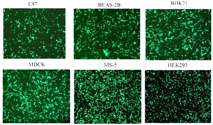
Figure 2: PolyMag Neo transfection efficiency in various cell lines.1x105 cells were transfected with 0.5 μg / well of pEGFP plasmid and 0.5 μL of PolyMag Neo reagent in 24-well plates. EGFP expression was monitored 24h after transfection by fluorescence microscopy.
Efforts to understand host factors critical for COVID-19 pathogenesis have identified high mobility group box 1 (HMGB1) to be crucial for regulating susceptibility to SARS-CoV-2. COVID-19 disease severity is correlated with heightened inflammatory responses, and HMGB1 is an important extracellular mediator in inflammation processes.In this study, we evaluated the effect of HMGB1 inhibitor Glycyrrhizin on the cellular perturbations in lung cells expressing SARS-CoV-2 viral proteins. Pyroptosis in lung cells transfected with SARS-CoV-2 S-RBD and Orf3a, was accompanied by elevation of IL-1β and extracellular HMGB1 levels. Glycyrrhizin mitigated viral proteins-induced lung cell pyroptosis and activation of macrophages. Heightened release of proinflammatory cytokines IL-1β, IL-6 and IL-8, as well as ferritin from macrophages cultured in conditioned media from lung cells expressing SARS-CoV-2 S-RBD and Orf3a was attenuated by glycyrrhizin. Importantly, Glycyrrhizin inhibited SARS-CoV-2 replication in Vero E6 cells without exhibiting cytotoxicity at high doses. The dual ability of Glycyrrhizin to concomitantly halt virus replication and dampen proinflammatory mediators might constitute a viable therapeutic option in patients with SARS-CoV-2 infection.
Alternative pre-mRNA splicing is key to proteome diversity; however, the biological roles of alternative splicing (AS) in signaling pathways remain elusive. Here, we focus on TEA domain transcription factor 1 (TEAD1), a YAP binding factor in the Hippo signaling pathway. Public database analyses showed that expression of YAP-TEAD target genes negatively correlated with the expression of a TEAD1 isoform lacking exon 6 (TEAD1ΔE6) but did not correlate with overall TEAD1 expression. We confirmed that the transcriptional activity and oncogenic properties of the full-length TEAD1 isoform were greater than those of TEAD1ΔE6, with the difference in transcription related to YAP interaction. Furthermore, we showed that RNA-binding Fox-1 homolog 2 (RBFOX2) promoted the inclusion of TEAD1 exon 6 via binding to the conserved GCAUG element in the downstream intron. These results suggest a regulatory mechanism of RBFOX2-mediated TEAD1 AS and provide insight into AS-specific modulation of signaling pathways.
Hinuma S., Vaccines (Basel) . 2022 Nov 30;10(12):2050.
(1) Background: Sodium taurocholate cotransporting polypeptide (NTCP) functions as a key receptor for the hepatitis B virus (HBV) infection. Analyzing HBV and NTCP interaction is an important issue not only for basic research but also for the development of anti-HBV therapeutics. We developed here a novel model system to analyze the interaction of NTCP with liposomes instead of HBV. (2) Methods: Liposomal binding and endocytosis through NTCP in HEK293T cells were achieved by serial treatments of HEL293T cells transiently expressing NTCP-green fluorescence protein (GFP) fusion protein with a synthetic biotinylated pre-S1 peptide (Myr47-Bio) and streptavidin (SA) complex (i.e., Myr47-Bio+SA) followed by biotinylated liposomes. By this procedure, binding of [biotinylated liposomes]-[Myr47-Bio+SA]-[NTCP-GFP] was formed. (3) Results: Using this model system, we found that liposomal binding to NTCP on the cell surface via Myr47-Bio+SA was far more efficient than that to scavenger receptor class B type 1 (SR-B1). Furthermore, liposomes bound to cell surface NTCP via Myr47-Bio+SA were endocytosed into cells after cells were cultured at 37 °C. However, this endocytosis was suppressed by 4 °C or cytochalasin B treatment. (4) Conclusions: This model system will be useful for not only analyzing HBV entry mechanisms but also screening substances to prevent HBV infection.
How proteins evolve new functionality is an important question in biology; prestin (SLC26A5) is a case in point. Prestin drives outer hair cell somatic motility and amplifies mechanical vibrations in the mammalian cochlea. The motility of mammalian prestin is analogous to piezoelectricity, in which charge transfer is coupled to changes in membrane area occupied by the protein. Intriguingly, nonmammalian prestin orthologs function as anion exchangers but are apparently nonmotile. We previously found that mammalian prestin is sensitive to membrane thickness, suggesting that prestin's extended conformation has a thinner hydrophobic height in the lipid bilayer. Because prestin-based motility is a mammalian specialization, we initially hypothesized that nonmotile prestin orthologs, while functioning as anion transporters, should be much less sensitive to membrane thickness. We found the exact opposite to be true. Chicken prestin was the most sensitive to thickness changes, displaying the largest shift in voltage dependence. Platypus prestin displayed an intermediate response to membrane thickness and gerbil prestin was the least sensitive. To explain these observations, we present a theory where force production, rather than displacement, was selected for the evolution of prestin as a piezoelectric membrane motor.
Valdez-Sinon AN., Star Protocols 2020 doi.org/10.1016/j.xpro.2020.100083
This protocol describes immunoprecipitation of proteins associated with FLAG-tagged recombinant proteins followed by mass spectrometry-based proteomics to identify the associated interactome components. FLAG epitope was chosen, because existing high-affinity monoclonal antibodies allow for sensitive immunoprecipitation and FLAG peptides permit efficient elution of protein complexes. With many commercially available FLAG tools, this protocol is highly versatile. This procedure reduces immunoprecipitation of nonspecific binding proteins. Gene ontology analyses performed following mass spectrometry-based proteomics may elucidate novel functions of proteins of interest.